Morningsunlight intensity modulates sleep spindle density via ipRGC‑driven thalamocortical calcium oscillations
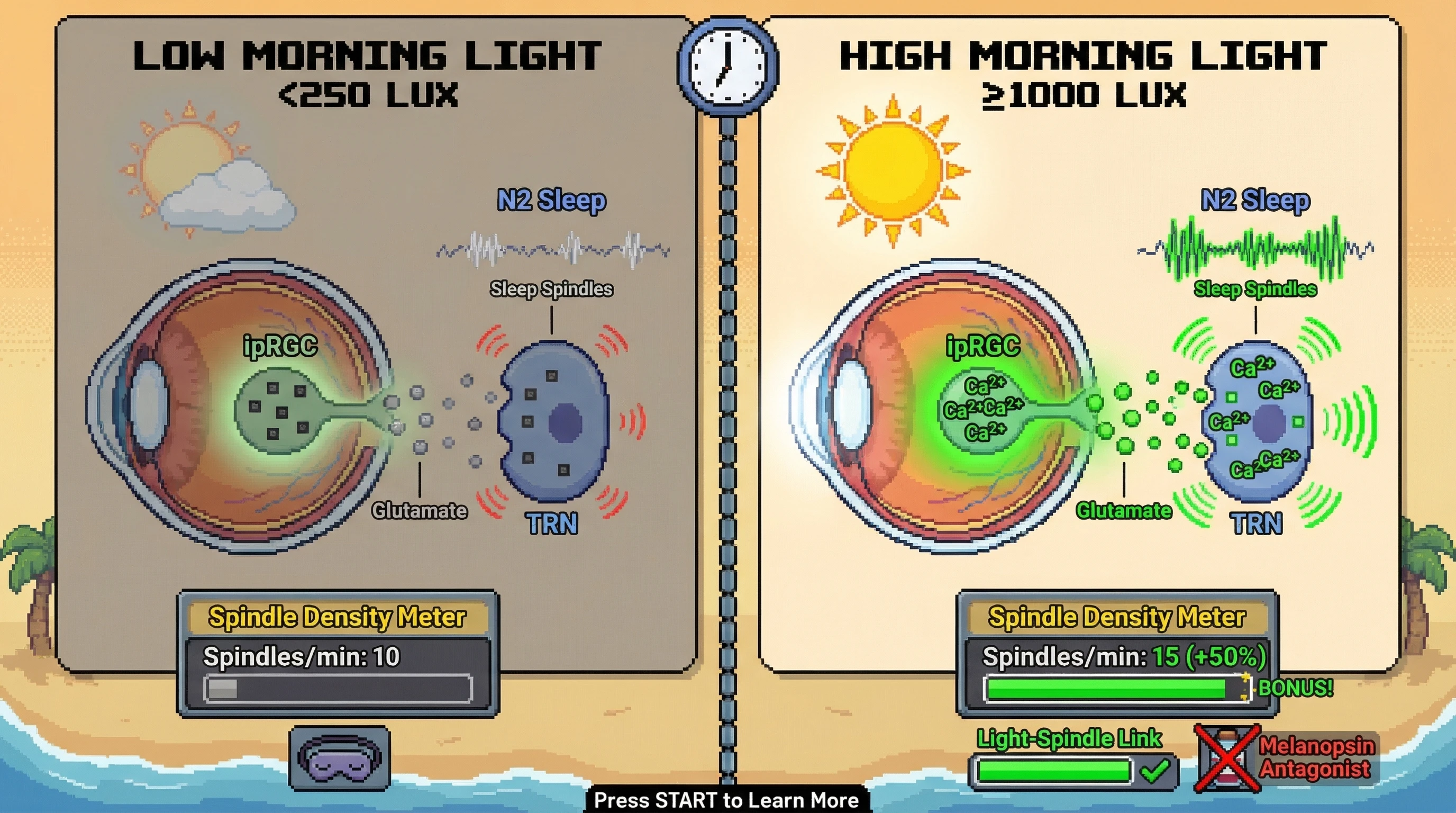
Hypothesis
Morning sunlight intensity, measured in lux, directly influences sleep spindle density through ipRGC‑mediated calcium signaling in the thalamic reticular nucleus.
Mechanistic rationale
- ipRGCs respond to blue‑green light and project to the suprachiasmatic nucleus, but collateral branches innervate the intergeniculate leaflet and thalamic regions.
- Light‑evoked melanopsin activation raises intracellular calcium in ipRGCs, which drives glutamatergic release onto thalamic reticular neurons.
- Elevated calcium in these GABAergic neurons enhances their burst firing, a key generator of sleep spindles during N2 sleep.
- Therefore, stronger morning light should produce a larger calcium imprint, increasing spindle propensity that night.
Testable predictions
- In healthy adults, a 30‑minute exposure to ≥1000 lux morning light will increase spindle density (spindles per minute of N2) by ≥15 % compared with <250 lux exposure.
- The relationship follows a saturating dose‑response curve, plateauing around 2000 lux.
- Blocking melanopsin signaling pharmacologically (e.g., with intravitreal antagonist) will abolish the light‑spindle link without affecting baseline spindle rates.
Experimental design (falsifiable)
- Crossover study, n = 30 participants, each undergoes four morning light conditions in random order: 0 lux (dim red <10 lux), 250 lux, 1000 lux, 2000 lux, all delivered via LED light boxes within 30‑60 min after waking for 30 min.
- Ambient temperature held at 18 °C, meal timing fixed, screen use prohibited after exposure.
- Polysomnography recorded the following night; spindle density, amplitude, and frequency extracted via automated detection (validated against expert scoring).
- Primary outcome: change in spindle density relative to baseline (0 lux).
- Null hypothesis: no significant differences across conditions.
- Rejection of null (p < 0.05, effect size >0.4) supports the hypothesis; failure to reject falsifies it.
Potential confounds and controls
- Individual differences in baseline melatonin amplitude will be measured via salivary dim‑light melatonin onset and entered as covariate.
- Sleep pressure controlled by keeping wake‑time constant and limiting naps.
- Retinal health screened to exclude ocular disease that could alter ipRGC function.
Broader implication
If confirmed, this would bridge the gap between circadian entrainment and micro‑architectural sleep benefits, offering a non‑pharmacological lever to boost spindle‑dependent memory consolidation and neuropsychiatric resilience.