Hypothesis: Peripheral nociceptor senescence drives systemic epigenetic aging via mitochondrial ROS spillover, making pain tolerance a causal readout of biological age
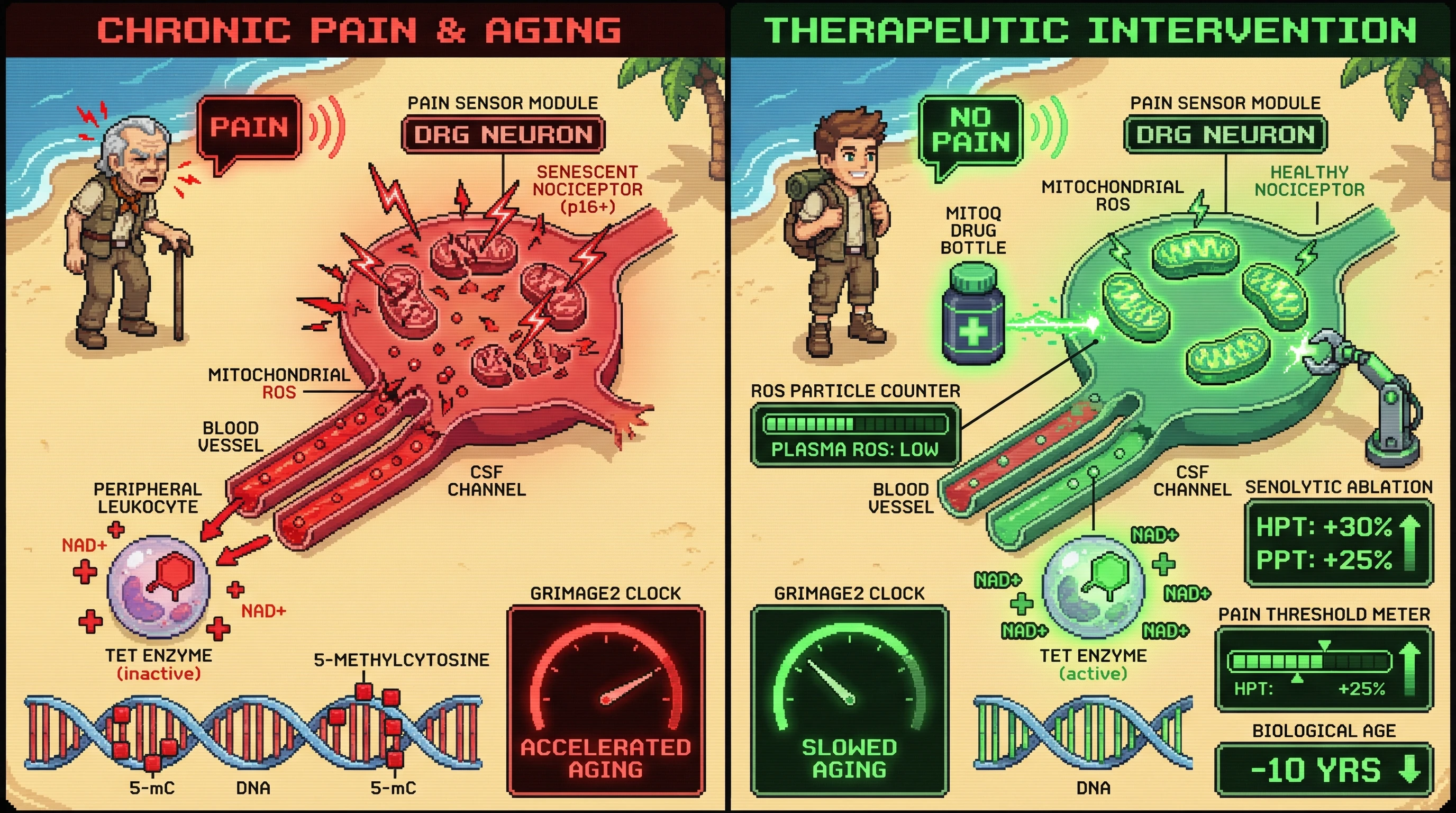
Hypothesis
Chronic pain sensitivity is not merely a biomarker but a driver of systemic aging through mitochondrial ROS release from senescent dorsal root ganglion (DRG) nociceptors, which accelerates epigenetic clocks in distant tissues.
Mechanistic Model
- Senescent nociceptor accumulation – With age or injury, a subset of DRG neurons expresses p16^INK4a^, p21, and SA‑β‑gal, adopting a senescence-associated secretory phenotype (SASP) that includes IL‑6, IL‑1β, and mitochondrial ROS (3, 4).
- Mitochondrial ROS spillover – Senescent DRG neurons exhibit fragmented mitochondria and elevated superoxide production. ROS can diffuse locally into cerebrospinal fluid and systemically via blood‑borne extracellular vesicles, oxidizing cytosolic NAD^+^ and impairing TET enzyme activity in peripheral leukocytes (5).
- Epigenetic age acceleration – Reduced TET activity leads to 5‑methylcytosine accumulation at GrimAge2 CpG sites, increasing predicted biological age independent of chronological age (1).
- Feedback loop – Elevated systemic ROS further sensitizes TRPV1 channels on remaining nociceptors, lowering pain thresholds and creating a vicious cycle of hypersensitivity and aging (6).
Testable Predictions
- Prediction 1: Selective ablation of senescent DRG neurons (using p16‑3MR transgenic mice + ganciclovir) will reduce mitochondrial ROS in plasma and slow GrimAge2 acceleration in liver and muscle, even without altering chronological age.
- Prediction 2: Administering a mitochondria‑targeted antioxidant (MitoQ) to aged wild‑type mice will normalize pain tolerance to youthful levels and decouple pain sensitivity from epigenetic age acceleration.
- Prediction 3: In humans, plasma cell‑free DNA bearing nociceptor‑specific methylation signatures (e.g., SCN10A promoter) will correlate positively with both heat pain threshold (HPT) decline and GrimAge2 acceleration.
Experimental Design
Animal Studies
- Groups: (i) Young (3 mo) WT, (ii) Aged (24 mo) WT, (iii) Aged WT + MitoQ, (iv) Aged p16‑3MR + ganciclovir, (v) Aged p16‑3MR vehicle.
- Measures: Baseline HPT and pressure pain threshold (PPT), plasma superoxide (DHE fluorescence), extracellular vesicle ROS content, GrimAge2 from liver/muscle DNA, circulating IL‑6.
- Analysis: Two‑way ANOVA with post‑hoc Tukey; mediation analysis to test whether ROS reduction mediates the effect of senescent cell removal on epigenetic age.
Human Pilot
- Cohort: 120 adults stratified by age (20‑35, 36‑55, 56‑75) and self‑reported chronic pain status.
- Procedures: Quantitative sensory testing (QST) for HPT and PPT, blood draw for plasma ROS, extracellular vesicle isolation, SCN10A‑methylated cfDNA via bisulfite sequencing, GrimAge2 calculation.
- Stats: Linear regression models testing cfDNA as mediator between QST outcomes and GrimAge2, adjusted for BMI, smoking, and medication.
Potential Confounds and Controls
- Systemic inflammation unrelated to nociceptors (e.g., periodontitis) will be screened and excluded via CRP.<br>
- Central sensitization contributions will be assessed via conditioned pain modulation (CPM) to ensure peripheral specificity.
- MitoQ off‑target effects will be controlled with a structurally similar inactive analog.
Implications
If validated, this hypothesis repositions pain tolerance as an actionable biomarker: interventions that clear senescent nociceptors or quench their mitochondrial ROS could simultaneously alleviate pain and retard systemic epigenetic aging. It also offers a non‑invasive, low‑cost readout (a 10‑minute QST battery) for tracking the efficacy of senolytic or mitochondrial therapies in clinical trials, addressing the current gap between molecular aging clocks and functional phenotype.