Ketosis-Primed Senolysis: Time-Restricted Eating Enhances Senolytic Efficacy via Autophagy-Dependent Mitochondrial Priming
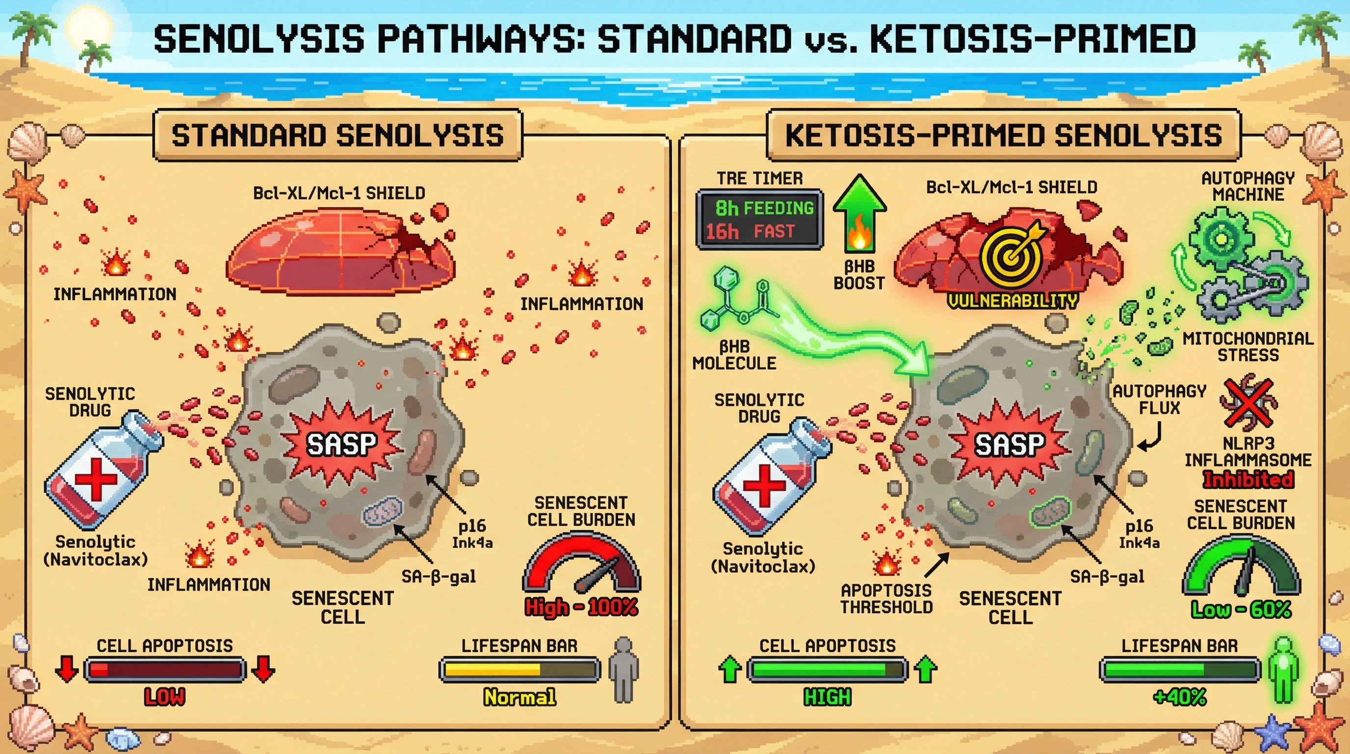
We hypothesize that administering senolytics during the peak of fasting-induced ketosis in a time-restricted eating (TRE) regimen will significantly increase senescent cell clearance compared with senolytic administration at random times, because ketone bodies (βHB) potentiate autophagy flux and remodel mitochondrial metabolism to create a synthetic lethal vulnerability in senescent cells.
Rationale: TRE induces autophagy flux and elevates circulating βHB, which has been shown to promote senolysis via βHB-mediated pathways in animal models [3]. Autophagy activation can directly trigger senolysis in certain senescent cell types [4], suggesting that autophagy‑primed senescent cells become more dependent on anti‑apoptotic Bcl‑2 family proteins for survival. Simultaneously, βHB inhibits NLRP3 inflammasome activity and alters NAD⁺/NADH ratios, shifting senescent cells toward a metabolic state that relies on heightened BCL‑XL or MCL‑1 expression to counteract increased pro‑apoptotic pressure from autophagy‑derived mitochondrial stress. Administering a Bcl‑2/xL inhibitor (e.g., navitoclax) or a Senolytic that targets these dependencies during the ketotic window should therefore push senescent cells over the apoptosis threshold more efficiently than dosing outside this window.
Experimental design: Use aged (20‑month) C57BL/6 mice divided into four groups (n=15 per group): 1) ad libitum feeding + vehicle; 2) ad libitum feeding + senolytic (navitoclax, 50 mg/kg i.p., weekly); 3) TRE (8‑hour feeding window, 16‑hour fast) + vehicle; 4) TRE + senolytic administered at ZT6 (mid‑fast, when βHB peaks, verified by tail‑bleed βHB measurement). Treatment lasts 12 weeks. Primary outcomes: senescent cell burden in liver, adipose, and kidney measured by p16^Ink4a^ immunofluorescence and SA‑β‑gal activity; secondary outcomes: autophagy flux (LC3A/LC3BII ratio, p62 degradation), circulating βHB, glucose tolerance, grip strength, and frailty index. Blood draws every 2 weeks to correlate senolytic timing with βHB peaks.
Predictions: Group 4 will show ≥40% greater reduction in senescent cell markers versus group 2, accompanied by heightened autophagy flux and improved metabolic phenotypes. If senolytic efficacy does not increase when timed to ketosis, the hypothesis is falsified. Additionally, if autophagy inhibition (via chloroquine co‑administration) abolishes the enhanced senolytic effect in group 4, it confirms autophagy as a necessary mediator.
Falsifiability: The hypothesis makes clear, quantitative predictions that can be tested with existing tools. A lack of significant interaction between TRE timing and senolytic potency, or failure to observe increased autophagy flux coinciding with βHB peaks, would refute the mechanistic link. Conversely, confirming the prediction would provide a mechanistic basis for optimizing senolytic schedules in humans, directly addressing the noted gap in combining TRE with senolytics [1,2,5].