Mitochondrial DNA–Derived Extracellular Vesicles from Senescent Cardiac Progenitors Propagate Senescence via cGAS‑STING Signaling in IGF‑1Rlow Niches
Hypothesis
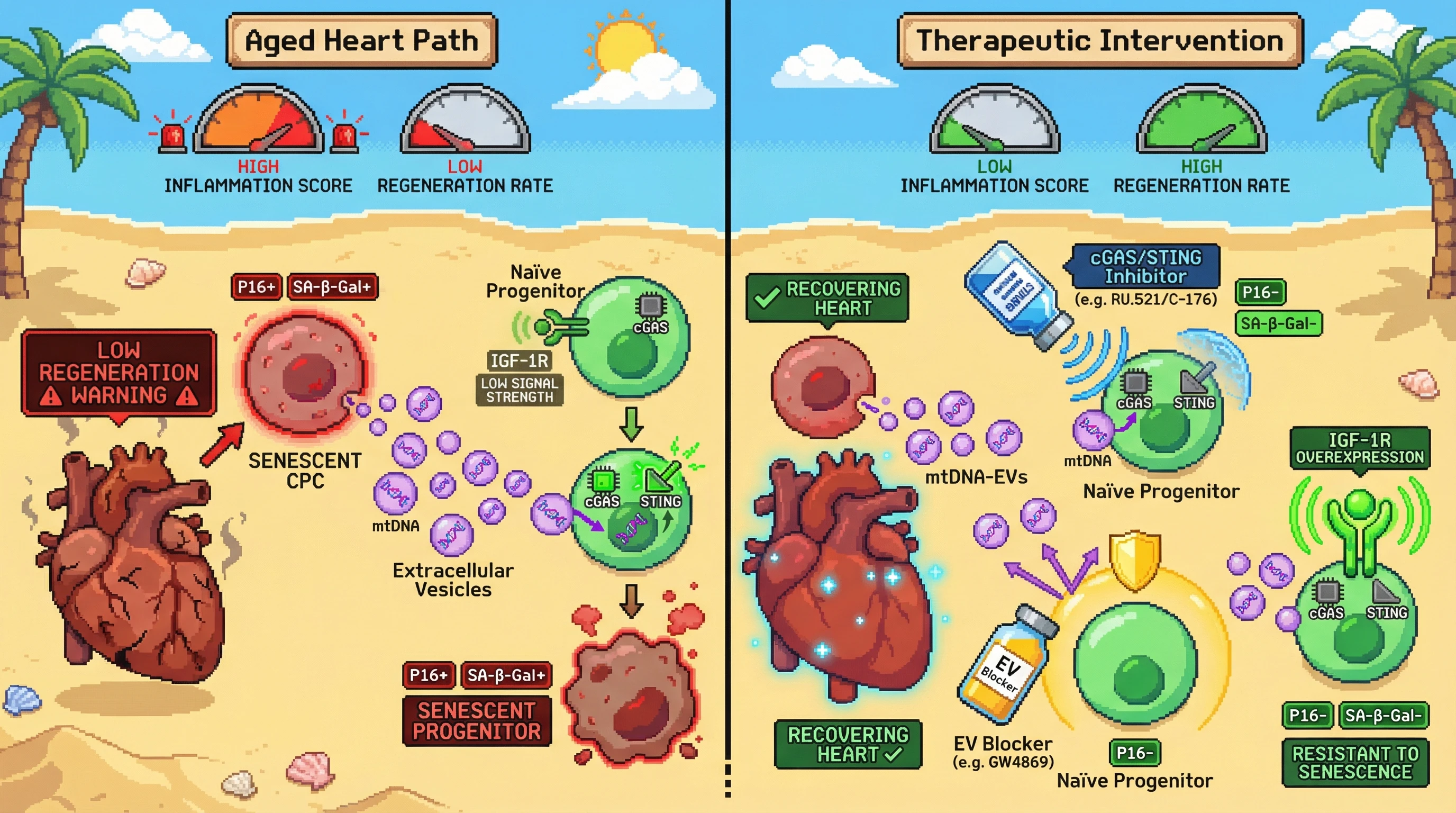
Senescent cardiac progenitor cells (CPCs) release mitochondrially‑derived DNA packaged in extracellular vesicles that activate the cGAS‑STING pathway in neighboring CD45⁻/c‑Kit⁺ progenitors, converting IGF‑1Rlow cells into a senescent state and amplifying the regenerative deficit in the aged heart.
Rationale
Aged hearts accumulate dysfunctional CPCs marked by p16^INK4A^, SA‑β‑gal, γH2AX and telomere attrition, which limits replication and differentiation [1]. These senescent CPCs secrete a SASP that induces paracrine senescence in adjacent cells [1]. Mitochondrial DNA integrity is required for CPC function, and age‑related mtDNA mutations impair proliferation [3]. Circulating cell‑free mtDNA (cf‑mt‑DNA) released from damaged cells can trigger innate immune sensors such as cGAS, leading to STING‑dependent IFN‑β production and senescence [4]. Moreover, only the CD45⁻/c‑Kit⁺ subpopulation exhibits robust clonogenic and myogenic capacity, while bulk c‑Kit⁺ cells are endothelial‑committed and poorly regenerative [2]. IGF‑1R marks a subset of human cardiac stem cells with superior therapeutic potential, yet its expression declines with age [5]. We propose that senescent CPCs selectively package damaged mtDNA into exosomes that are taken up by IGF‑1Rlow CD45⁻/c‑Kit⁺ progenitors, where mtDNA engages cGAS, activating STING and driving a senescent phenotype that suppresses IGF‑1R signaling and regenerative output.
Testable Predictions
- Isolates of extracellular vesicles from senescent CPCs will contain elevated mtDNA relative to vesicles from young CPCs.
- Exposure of naïve CD45⁻/c‑Kit⁺ progenitors to senescent‑CPC vesicles will increase cGAS activation, STING phosphorylation, IFN‑β secretion, and acquisition of p16^INK4A^ and SA‑β‑gal positivity.
- Blocking mtDNA uptake (e.g., with exosome synthesis inhibitor GW4869) or inhibiting cGAS/STING (using small‑molecule antagonists) will prevent vesicle‑induced senescence in CD45⁻/c‑Kit⁺ cells.
- In aged mouse hearts, genetic deletion of cGAS in CD45⁻/c‑Kit⁺ cells or pharmacological STING inhibition will reduce senescence markers among progenitors, improve IGF‑1R signaling, and enhance post‑injury cardiomyocyte renewal.
- Overexpression of IGF‑1R in CD45⁻/c‑Kit⁺ progenitors will rescue resistance to vesicle‑mediated senescence, even in the presence of high cf‑mt‑DNA levels.
Experimental Approach
- Cell models: isolate cardiac progenitor cells from young (3‑month) and aged (24‑month) mice; sort CD45⁻/c‑Kit⁺ and IGF‑1Rhigh/low fractions using flow cytometry.
- Senescence induction: treat young CPCs with low‑dose doxorubicin or irradiation to generate a senescent phenotype validated by p16^INK4A^, SA‑β‑gal, γH2AX.
- Vesicle purification: collect conditioned media, isolate exosomes via ultracentrifugation or size‑exclusion chromatography; quantify mtDNA by qPCR for mt‑ND1 normalized to vesicle protein.
- Uptake assay: label vesicles with PKH26, incubate with naïve CD45⁻/c‑Kit⁺ IGF‑1Rlow cells, confirm internalization by confocal microscopy.
- Signaling readouts: measure cGAS binding (DNA‑pull down), STING phosphorylation (Western blot), IFN‑β ELISA, and downstream ISG expression (qPCR for MX1, OAS1).
- Senescence assessment: after 48‑72 h vesicle exposure, evaluate p16^INK4A^, SA‑β‑gal, EdU incorporation, and mitochondrial membrane potential (JC‑1).
- Intervention tests: apply GW4869 (exosome release inhibitor), cGAS inhibitor RU.521, or STING antagonist C‑176; assess rescue of proliferation and reduction of SASP cytokines (IL‑6, IL‑8) via Luminex.
- In vivo validation: generate aged mice with CD45⁻/c‑Kit⁺‑specific cGAS knockout (using cKit‑CreER) or treat with STING inhibitor; after myocardial infarction, quantify fibrosis (Masson’s trichrome), new cardiomyocytes (α‑actinin^+ Ki67^+), and IGF‑1R activity (p‑AKT).
- Rescue experiment: transduce IGF‑1Rlow progenitors with IGF‑1R‑overexpressing lentivirus prior to vesicle challenge; assess whether senescence markers are blunted.