Sleep‑Dependent Lysosomal Reboot: CSTB‑Mediated Cathepsin Control as the Glymphatic Autopsy Mechanism
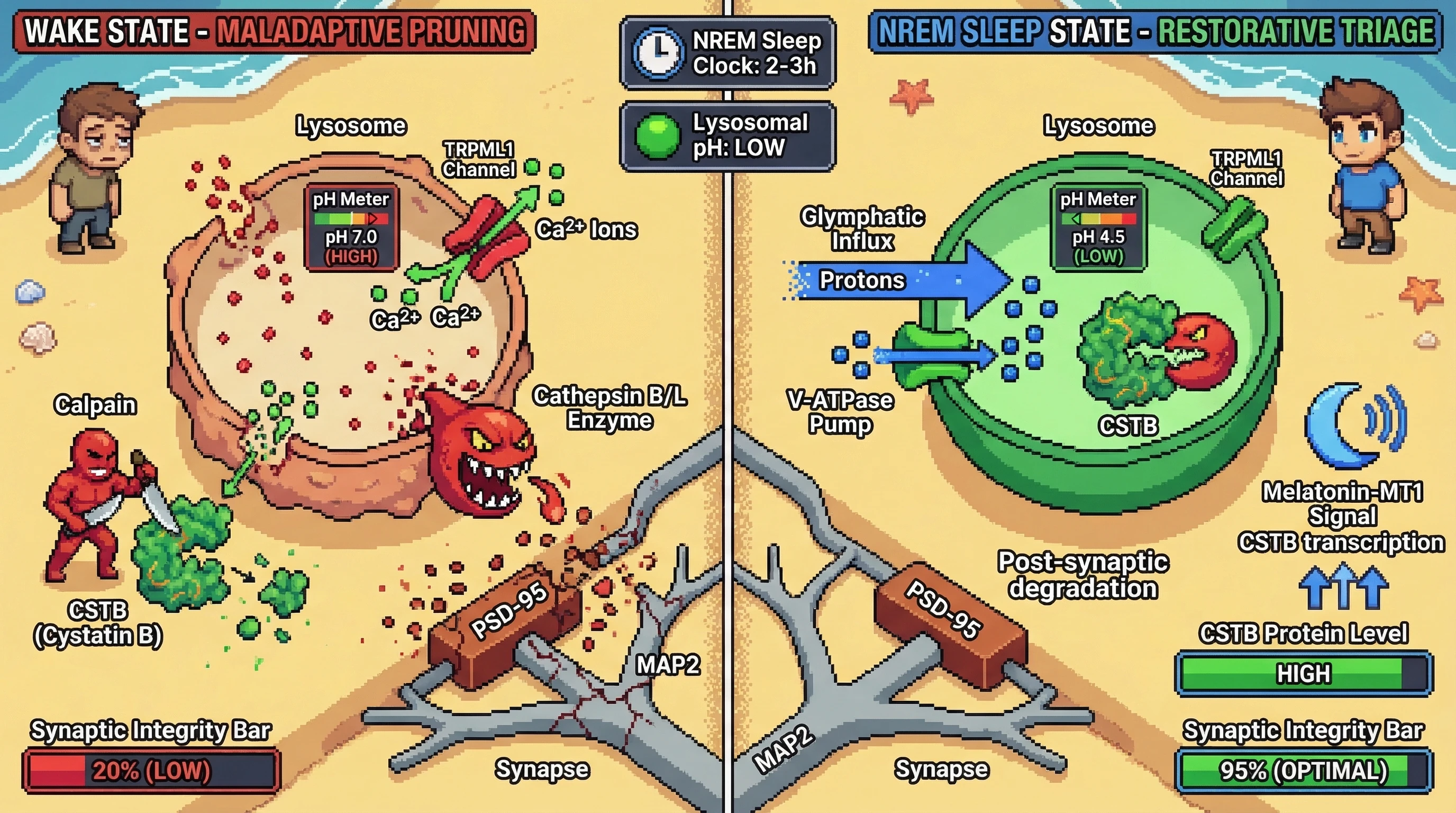
Hypothesis
During NREM slow‑wave sleep the brain actively re‑acidifies lysosomes and upregulates cystatin B, converting the glymphatic influx into a controlled proteolytic triage that trims excess synapses; sleep loss blocks this reset, allowing cathepsin leakage to drive maladaptive synaptic pruning.
Mechanistic Basis
- Aging lysosomes lose membrane integrity, raising pH and triggering TRPML1‑mediated Ca2+ release that activates calpains, further damaging the membrane【https://pmc.ncbi.nlm.nih.gov/articles/PMC7388076/】.
- Cytosolic Ca2+ also activates calpains that cleave CSTB, diminishing its inhibitory potency on cathepsins B and L【https://pmc.ncbi.nlm.nih.gov/articles/PMC11753708/】.
- CSTB normally restrains chromatin‑associated cathepsin L, preventing aberrant histone degradation【https://www.frontiersin.org/journals/molecular-neuroscience/articles/10.3389/fnmol.2022.1069122/full】.
- Slow‑wave sleep coincides with a surge in glymphatic CSF‑interstitial exchange that delivers protons and ATP‑driven V‑ATPase activity to lysosomes, re‑acidifying them and inhibiting TRPML1 opening【https://pmc.ncbi.nlm.nih.gov/articles/PMC8202514/】.
- Concurrently, sleep‑dependent signaling (e.g., via melatonin‑MT1 receptors) boosts CSTB transcription, reinforcing the cathepsin brake【https://pubmed.ncbi.nlm.nih.gov/41271184/】.
- When this window is missed, leaked cathepsins degrade PSD‑95, MAP2 and other synaptic scaffolds, producing the synaptic loss seen in chronic sleep disruption.
Testable Predictions
- Cortical lysosome pH will be lowest during early NREM and rise progressively with wakefulness.
- CSTB protein levels will peak after 2–3 h of consolidated sleep and trough after prolonged wakefulness.
- Pharmacological blockade of TRPML1 or genetic overexpression of CSTB in mice will prevent sleep‑deprivation‑induced cathepsin B/L activity and preserve synaptic protein levels.
- Conversely, chemogenetic inhibition of V‑ATPase during sleep will mimic the lysosomal pH rise seen in wakefulness and exacerbate cathepsin‑mediated synaptic degradation.
Experimental Approach
- Record in vivo lysosomal pH using LysoSensor‑yellow/blue dye in Thy1‑LysoSensor mice across sleep‑wake states measured by EEG/EMG.
- Quantify CSTB and cathepsin activity via western blot and activity‑based probes in microdissected cortex at ZT2 (early sleep) vs ZT14 (late wake).
- Subject mice to 6 h sleep deprivation; treat subsets with ML‑SI1 (TRPML1 inhibitor) or AAV‑CSTB; assess cathepsin leakage (immunostaining for cytosolic cathepsin L) and dendritic spine density (DiOlistic labeling).
- Rescue experiments: administer cathepsin B/L inhibitor (Ca‑074Me) during deprivation to test whether synaptic loss is protease‑dependent.
- Statistical plan: two‑way ANOVA (state × treatment) with post‑hoc Tukey; n ≥ 8 per group for power > 0.8.
If predictions hold, sleep’s autopsy function is recast as a lysosomal quality‑control step that gates which neural architectures survive to the next day. Failure of this gate—through impaired re‑acidification or CSTB loss—converts a restorative process into a source of neurodegeneration.