Heparan sulfate proteoglycan loss exposes cryptic collagen IV sites that drive alternative complement pathway activation and local C3 synthesis in retinal aging
Hypothesis
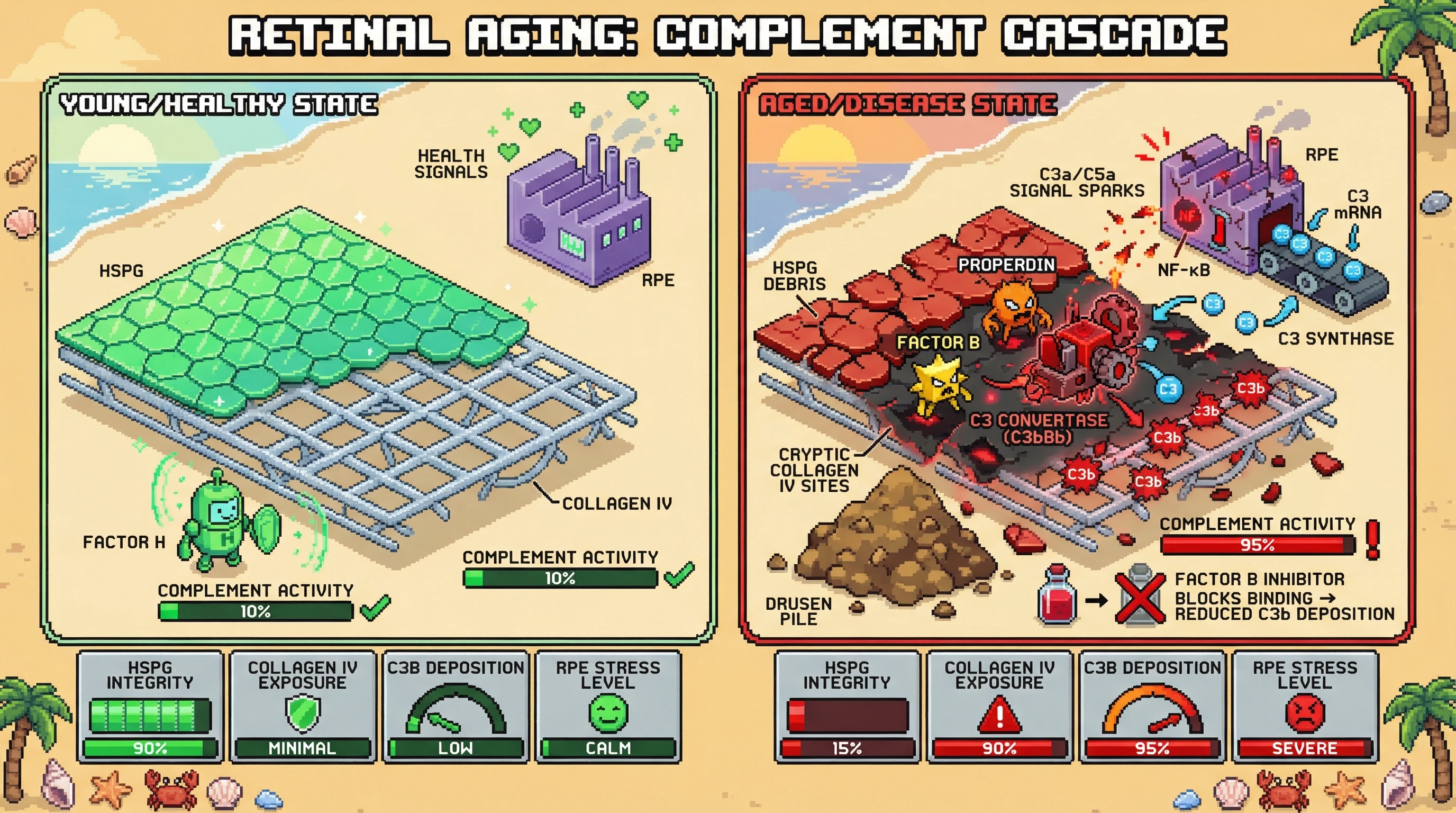
Loss of heparan sulfate proteoglycans (HSPGs) in aging Bruch's membrane not only diminishes Factor H binding but also unmasks cryptic binding sites on collagen IV that directly initiate the alternative complement pathway. These exposed sites recruit Factor B and properdin, forming C3 convertase (C3bBb) independent of classical or lectin routes. The resulting C3b deposition amplifies local C3 synthesis in retinal pigment epithelium (RPE) via a complement‑triggered NF‑κB loop, creating a self‑reinforcing micro‑environment that drives drusen formation and RPE stress.
Mechanistic Reasoning
- HSPG loss reduces steric hindrance and alters electrostatic shielding of collagen IV fibrils.
- Exposed collagen IV domains (e.g., the COL1 region) possess high affinity for Factor B, as shown in extracellular matrix models.
- Factor B binding stabilizes the C3bBb convertase, leading to sustained C3 cleavage and C3b amplification.
- Elevated C3a and C5a engage receptors on RPE, activating NF‑κB and upregulating C3 transcription.
- Increased intracellular C3 feeds back to amplify complement activation at the RPE‑Bruch's interface, establishing a vicious cycle.
Testable Predictions
- Prediction 1: In aged mouse eyes or human donor Bruch's membrane with validated HSPG loss, collagen IV fragments will co‑immunoprecipitate with Factor B and properdin more intensely than in young tissue.
- Prediction 2: Enzymatic restoration of HSPG sulfation using heparan sulfate mimetics will decrease collagen IV–Factor B interaction and lower C3b deposition in ex‑vivo choroid‑RPE explants.
- Prediction 3: Knock‑down of collagen IV in RPE cultures will attenuate Factor B recruitment and reduce NF‑κB‑driven C3 mRNA expression despite HSPG deficiency.
- Prediction 4: Pharmacological inhibition of Factor B (e.g., with a small‑molecule antagonist) will block the increase in local C3 synthesis seen in HSPG‑deficient RPE, whereas inhibition of classical pathway components (C1q) will have minimal effect.
Experimental Approaches
- Perform immunoblot and proximity ligation assays on Bruch's membrane extracts from young vs. aged donors to detect collagen IV–Factor B complexes.
- Treat explant cultures with synthetic heparan sulfate (e.g., pi‑88) and quantify C3b deposition by immunofluorescence and ELISA.
- Use siRNA against collagen IV in primary human RPE cells cultured on HSPG‑depleted matrices; measure C3 mRNA (qPCR) and secreted C3 protein.
- Apply Factor B inhibitor (e.g., BMS‑986168) or C1q blocking antibodies; assess downstream C3 activation and NF‑κB nuclear translocation.
Falsifiability
If collagen IV–Factor B interaction does not increase with HSPG loss, or if restoring HSPG sulfation fails to reduce C3b deposition, the proposed mechanism would be refuted. Similarly, if Factor B inhibition does not diminish local C3 synthesis in HSPG‑deficient RPE, the hypothesis would be unsupported.
Linking structural ECM alterations to a direct alternative‑pathway trigger provides an upstream target that complements existing C3/C5 therapies and may allow early intervention before irreversible retinal damage.