Activity-dependentAPE1 dephosphorylation creates a BER lyase bottleneck that drives toxic abasic site accumulation in aging neurons
Hypothesis
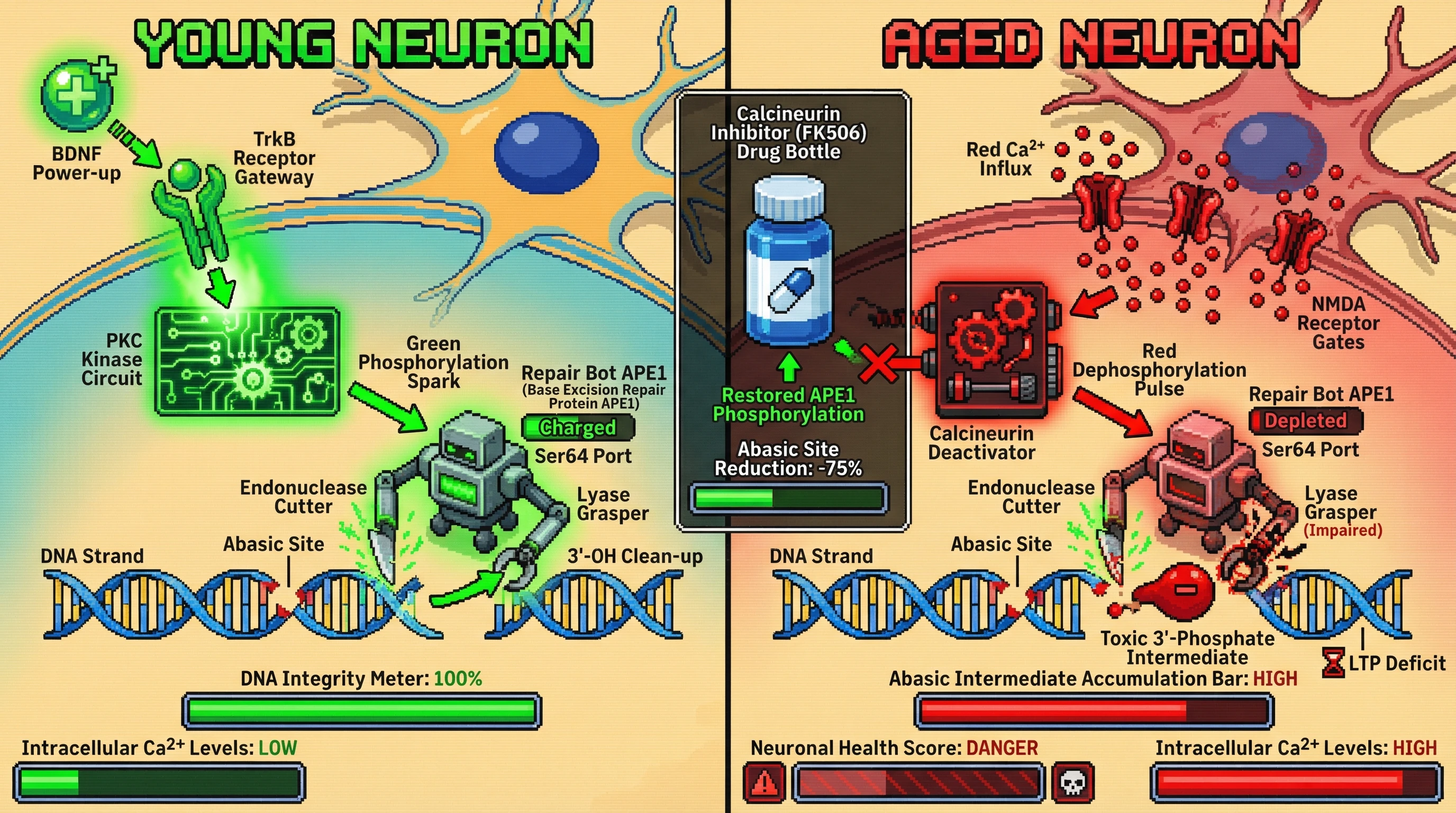
Activity-dependent calcium influx in aging neurons triggers calcineurin-mediated dephosphorylation of APE1 at a specific serine residue, selectively impairing its AP-lyase activity while leaving AP endonuclease function intact. This creates a kinetic bottleneck at the lyase step, causing accumulation of toxic 3′-phosphate/aldehyde abasic intermediates that are more deleterious than unrepaired 8-oxoG.
Mechanistic Basis
- BDNF–TrkB signaling normally sustains APE1 phosphorylation via the PLCγ–PKC cascade (1). Age-related decline in BDNF reduces this kinase activity, shifting the balance toward phosphatase dominance.
- Elevated intracellular Ca²⁺ from dysregulated NMDA receptors activates calcineurin, which dephosphorylates APE1 (predicted site Ser⁶⁴ based on consensus motifs). Dephosphorylated APE1 retains AP endonuclease activity but shows reduced β‑elimination/lyase activity, as shown for other APE1 phospho‑mutants (3).
- The resulting AP‑site intermediate accumulates because POLβ recruitment requires a properly processed 3′ terminus; the blocked lyase step leaves a 3′‑phosphate that inhibits POLβ, leading to persistent, potentially mutagenic intermediates (4).
Testable Predictions
- Phospho‑specific immunoblotting of neuronal extracts from young vs. aged mice will show decreased p-APE1 (Ser⁶⁴) correlating with increased 3′-phosphate abasic sites measured by the aldehyde‑reactive probe assay.
- Pharmacological inhibition of calcineurin (e.g., FK506) in aged neuron cultures will restore APE1 phosphorylation, reduce abasic intermediate levels, and rescue LTP deficits.
- Expression of phospho‑mimetic APE1 (S⁶⁴D) in Ogg1‑deficient neurons will ameliorate toxic intermediate accumulation and improve survival despite low OGG1.
- BDNF supplementation will rescue APE1 phosphorylation and reduce intermediate accumulation in an TrkB‑dependent manner, blocked by K252a.
Potential Implications
If validated, this model shifts focus from merely boosting OGG1 activity to preserving APE1 lyase function through phosphatase inhibition or BDNF‑mimetic strategies. It also explains why mitochondrial OGG1 overexpression improves cognition (2): mitochondrial BER may be less affected by calcium‑calcineurin signaling, highlighting compartment‑specific regulation.