Estrogen loss drives a fibroblast sulfotransferase switch that reshapes dermal chondroitin sulfate sulfation and exports a fibrosis-predictive exosomal GAG signature
Hypothesis
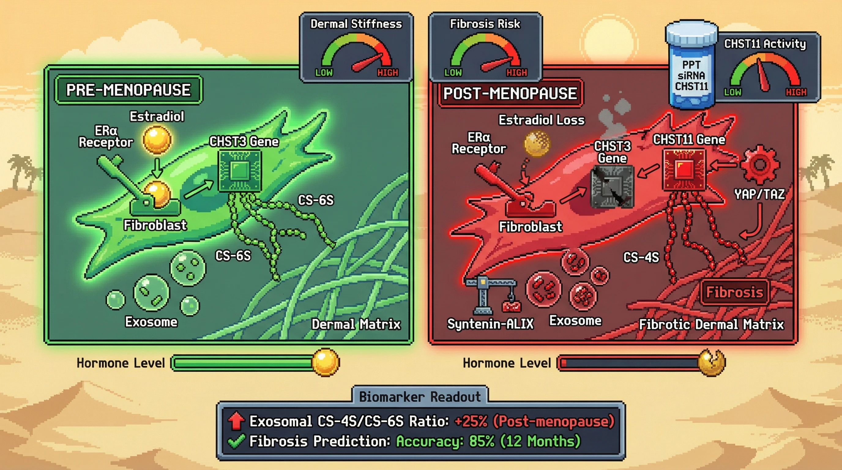
Estrogen loss in perimenopausal fibroblasts shifts chondroitin sulfate sulfation from the 6‑O‑ to the 4‑O‑position, and the altered GAG motif is packaged into exosomes that circulate as a predictive signature of impending dermal fibrosis.
Mechanistic Basis
Estrogen receptor α (ERα) directly binds promoter regions of the chondroitin‑6‑sulfotransferase (CHST3) gene, sustaining its expression in pre‑menopausal fibroblasts [GlycoAgeTest]. When estradiol declines, ERα activity falls, CHST3 transcription drops while the 4‑O‑sulfotransferase (CHST11) is derepressed via loss of ER‑mediated repression [Developmental GAG regulation]. The resulting CS‑4S enrichment increases electrostatic binding to collagen fibrils, promoting fibrillar alignment and matrix stiffening [ECM structural memory]. Stiff matrices activate fibroblast mechanotransduction (YAP/TAZ), which further upregulates CHST11, creating a feed‑forward loop. Altered CS‑4S chains are sorted into exosomes through interaction with syntenin‑ALIX cargo adapters, yielding EV‑borne GAG profiles that mirror the dermal ECM state.
Testable Predictions
- In skin biopsies from women aged 40‑55, CS‑4S/CS‑6S ratios will rise sharply after the onset of menopause and correlate with serum estradiol levels (inverse relationship).
- Exosomes isolated from peripheral blood of the same cohort will show a matching increase in CS‑4S‑containing chondroitin sulfate chains, and this exosomal GAG ratio will predict an increase in dermal stiffness measured by cutometry ≥12 months later.
- Pharmacological activation of ERα (e.g., with selective agonist PPT) in cultured human dermal fibroblasts will rescue CHST3 expression, reduce CS‑4S/CS‑6S ratio, and decrease exosomal loading of CS‑4S chains.
- Knock‑down of CHST11 in fibroblasts from post‑menopausal donors will normalize exosomal CS‑4S levels and attenuate TGF‑β‑induced collagen I secretion.
Experimental Approach
- Collect paired skin punch biopsies and blood from pre‑menopausal (n=30), perimenopausal (n=30), and post‑menopausal (n=30) women; quantify CS‑4S and CS‑6S by DSA‑FACE HPLC‑FLR/MS [GlycoAgeTest].
- Isolate exosomes via ultracentrifugation, perform LC‑MS/MS‑based GAG sequencing to determine sulfation patterns.
- Measure dermal stiffness using a cutometer; follow participants longitudinally for 18 months to record incidence of clinically diagnosed skin fibrosis (e.g., scleroderma‑like changes).
- In vitro, treat primary fibroblasts with estradiol, PPT, or siRNA against CHST11/CHST3; assess CHST mRNA (qPCR), sulfotransferase activity (radiometric assay), and exosomal GAG cargo.
- Use luciferase reporters containing CHST3 or CHST11 promoters to test ERα binding (ChIP‑qPCR). Statistical analysis: linear mixed models for biomarker trajectories, ROC curves for predictive power, and interaction terms for sex and menopausal status.
If the predictions hold, the hypothesis establishes a mechanistic link between hormonal decline, ECM sulfation remodeling, and exosomal communication that could be intercepted with ERα‑targeted therapies or exosome‑based biomarkers.