Pericyte Loss‑Driven Zonal Hypoxia Explains Selective DHEA Decline and Cortisol Rise in Aging Adrenal Cortex
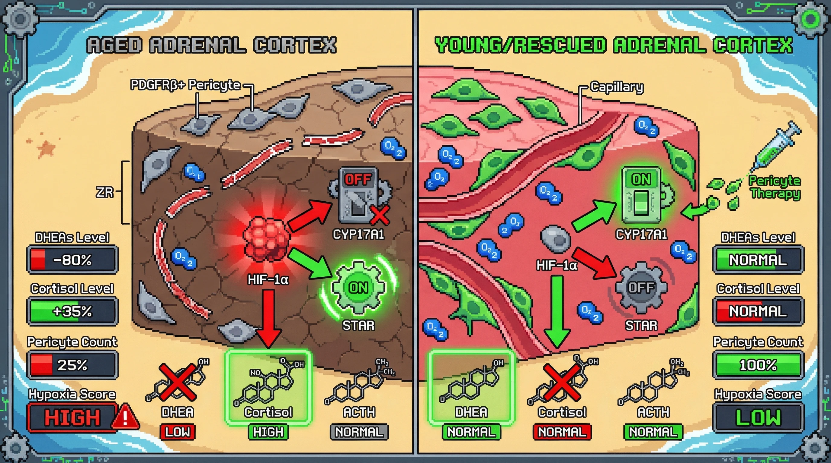
Hypothesis
Age‑related loss of adrenal cortical pericytes triggers capillary rarefaction specifically in the zona reticularis, creating a hypoxic microenvironment that suppresses DHEA synthesis via HIF‑1α‑mediated inhibition of CYP17A1 while simultaneously enhancing cortisol output through StAR upregulation and altered mitochondrial redox state.
Mechanistic Basis
- Pericytes express PDGFRβ and NG2; their depletion reduces VEGF‑mediated angiogenic support, leading to endothelial dropout and impaired oxygen delivery.
- Hypoxia stabilizes HIF‑1α, which binds to the promoter of CYP17A1 and represses transcription, lowering DHEA precursor flux.
- Concurrently, HIF‑1α upregulates STAR and increases mitochondrial ROS, favoring pregnenolone shunting toward the glucocorticoid pathway.
- This dual effect explains the observed 5‑10‑fold DHEAS decline (adrenopause) and the 20‑50% cortisol rise without ACTH change.
- The flattened cortisol awakening response follows from reduced adrenal reserve and impaired rapid steroidogenic burst.
Testable Predictions
- Aged mice will show a >40% reduction in PDGFRβ⁺ pericytes confined to the zona reticularis, whereas zona glomerulosa and fasciculata remain relatively spared.
- Pharmacologic stabilization of HIF‑1α (e.g., using DMOG) in young adrenal slices will reproduce the aged steroid profile: decreased DHEA, increased cortisol.
- Pericyte replacement via intradrenal injection of PDGFRβ⁺ progenitors will restore DHEAS secretion and normalize the cortisol awakening rhythm in aged animals.
- In humans, circulating soluble PDGFRβ levels will inversely correlate with DHEAS and directly with evening cortisol, independent of ACTH.
Experimental Approach
- Use adrenal zona‑specific Cre lines (e.g., CYP11B2‑Cre for glomerulosa, CYP11B1‑Cre for fasciculata, and HSD3B2‑Cre for reticularis) crossed with PDGFRβ‑flux reporters to quantify pericyte density across the lifespan.
- Apply pimonidazole hypoxia adduct staining and HIF‑1α immunohistochemistry to map oxygen tension.
- Measure steroid outputs via LC‑MS/MS before and after pericyte depletion (using diphtheria toxin‑mediated ablation) or rescue.
- Validate in a cohort of older adults: plasma soluble PDGFRβ, DHEAS, cortisol, and ACTH, plus MRI‑derived adrenal perfusion.
Implications
If confirmed, this mechanism positions pericyte health as a upstream regulator of adrenopause and offers a translational target: pericyte‑supportive therapies (e.g., angiopoietin‑1 mimetics) could mitigate age‑related HPA dysregulation and its downstream cognitive and metabolic risks.
Citations
- et al. 2019: adrenal cortex aging and stress response 1
- Liu et al. 2023: cortisol awakening response and cognition 2
- McEwen 2017: cortisol effects on hippocampus 3
- Lupien et al. 2014: HPA dysfunction and depression 4
- Evans et al. 2012: CAR and executive function 5
- Russel et al. 2023: individual variability in HPA axis 6
- Dickman 2025: bioprinted adrenal tissues 7
- Recent hypotheses on adrenal vasculature 8
- DHEA decline discussion 9
- Watershed ischemia hypothesis 10