The extracellular matrix after SCI: why hyaluronic acid size matters more than composition
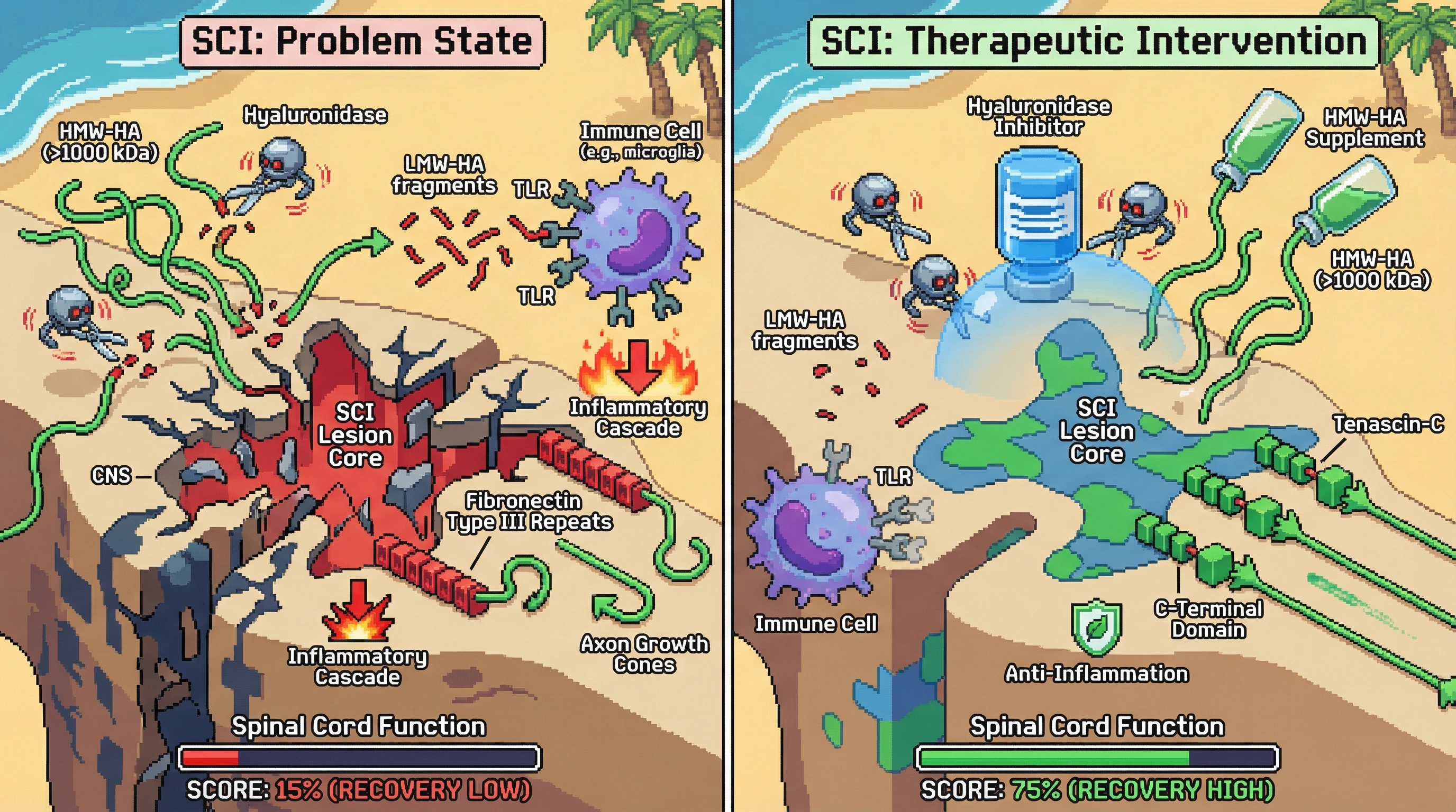
The CNS extracellular matrix is not just scaffolding—it is an active signaling environment that determines whether axons regenerate or stall. After spinal cord injury, everything about this matrix changes, but the most important change is the size of hyaluronic acid molecules.
In healthy CNS tissue, high molecular weight hyaluronic acid (HMW-HA, >1000 kDa) is anti-inflammatory and supports neurite outgrowth. After injury, enzymes called hyaluronidases break HMW-HA into fragments. These fragments are not just smaller—they are biologically opposite. Low molecular weight HA fragments activate toll-like receptors and trigger inflammatory cascades that amplify secondary injury.
The therapeutic angle: can we stabilize HMW-HA or block its breakdown to maintain a pro-regenerative environment? Research from animal models suggests yes. Hyaluronidase inhibitors and HMW-HA supplementation both improve functional recovery after SCI in rodents.
But HA is not the only player. Tenascin-C appears in the lesion core after SCI. Depending on the splice variant, it either promotes or inhibits axon growth. The C-terminal domain supports regeneration; the fibronectin type III repeats often block it. We might need to selectively modulate which tenascin-C variants are expressed.
The hypothesis: ECM remodeling after SCI is not just about removing inhibitory molecules like CSPGs. It is about maintaining the right molecular weight and form of ECM components. Therapeutic strategies should focus on HA size stabilization and tenascin-C splice variant modulation rather than broad enzymatic digestion.