IFN-I reprogramstissue macrophages via PI3K-AKT-Foxo1 to amplify SASP and drive inflammaging
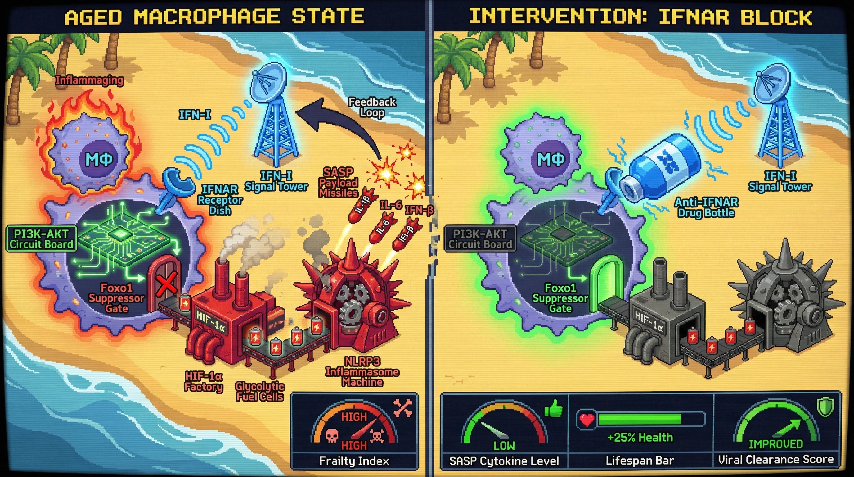
Hypothesis
Chronic low‑grade IFN‑I signaling reprograms tissue‑resident macrophages through sustained PI3K‑AKT activation and Foxo1 inhibition, shifting them toward a glycolytic, HIF‑1α‑driven state that amplifies the SASP via NLRP3 inflammasome activation and creates a self‑reinforcing IFN‑I/SASP loop that drives inflammaging.
Mechanistic Rationale
- In γ/δ T cells, tonic IFN‑I downregulates Foxo1 via PI3K‑AKT, expanding IL‑17‑producing subsets 1.
- Similar PI3K‑AKT‑Foxo1 signaling controls macrophage metabolism; Foxo1 loss promotes HIF‑1α stabilization and glycolytic shift, which is known to prime NLRP3 inflammasome activity.
- Active NLRP3 releases IL‑1β and caspase‑1‑processed gasdermin D, prompting NF‑κB‑dependent transcription of SASP cytokines (IL‑6, IL‑8, S100A8/A9) and IFN‑β, thereby feeding back onto IFNAR in an autocrine fashion.
- This loop explains the observed STAT3 bias in monocytes and CD4⁺ T cells 2, as SASP‑derived IFN‑I favors STAT3 activation while inducing SOCS3‑mediated STAT1 suppression.
- The metabolic shift also links to progeroid phenotypes 3 and accounts for the paradox of heightened IFN‑I tone with reduced antiviral responsiveness via chronic STING overstimulation 4.
Testable Predictions
- Blocking IFNAR specifically in macrophages (e.g., using LysM‑Cre‑Ifnar1 floxed mice) will reduce HIF‑1α target genes, NLRP3 inflammasome activation, and SASP secretion in aged tissues.
- Genetic deletion of Foxo1 in macrophages will phenocopy the aged SASP profile even in young mice, whereas macrophage‑specific Foxo1 overexpression will suppress SASP despite systemic IFN‑I elevation.
- Pharmacologic inhibition of PI3K‑AKT (e.g., with a selective AKT inhibitor) in aged mice will normalize STAT3/STAT1 balance in monocytes and improve viral clearance without abolishing baseline IFN‑I tone.
- In humans, ex vivo treatment of peripheral blood monocytes from older donors with an IFNAR antagonist will decrease phospho‑S6 (PI3K‑AKT readout) and HIF‑1α, correlating with reduced IL‑1β and S100A8/A9 production.
Experimental Approach
- Generate macrophage‑specific Ifnar1 and Foxo1 conditional KO mice; assess aging‑related phenotypes (frailty index, tissue histology, serum SASP cytokines) and challenge with influenza virus to measure antiviral immunity.
- Perform flow cytometry and phospho‑flow on sorted macrophages from young vs. old mice to quantify p‑AKT, Foxo1 nuclear exclusion, HIF‑1α, and cleaved caspase‑1.
- Use Seahorse glycolysis assays to link IFN‑I tone to metabolic reprogramming.
- In parallel, collect monocytes from a longitudinal cohort of healthy adults (ages 20‑80); treat ex vivo with anti‑IFNAR2 antibody and measure changes in p‑STAT1/STAT3, SOCS3, and SASP mediators by multiplex ELISA and RNA‑seq.
- Computational modeling: integrate phospho‑proteomics, transcriptomics, and metabolomics data to simulate the IFN‑I→PI3K‑AKT→Foxo1→HIF‑1α→NLRP3→SASP→IFN‑I circuit and predict intervention points.
If validated, this hypothesis would reposition macrophage‑intrinsic PI3K‑AKT‑Foxo1 signaling as a central node linking interferonopathy to inflammaging, offering a tractable target for breaking the IFN‑I/SASP feedback loop while preserving host defense.