Corneal Confocal Microscopy Nerve Fiber Density Decline Rate Predicts Systemic Small Fiber Neuropathy Progression in Primary Sjögren Syndrome 6–12 Months Before Quantitative Sensory Testing Deterioration
Background
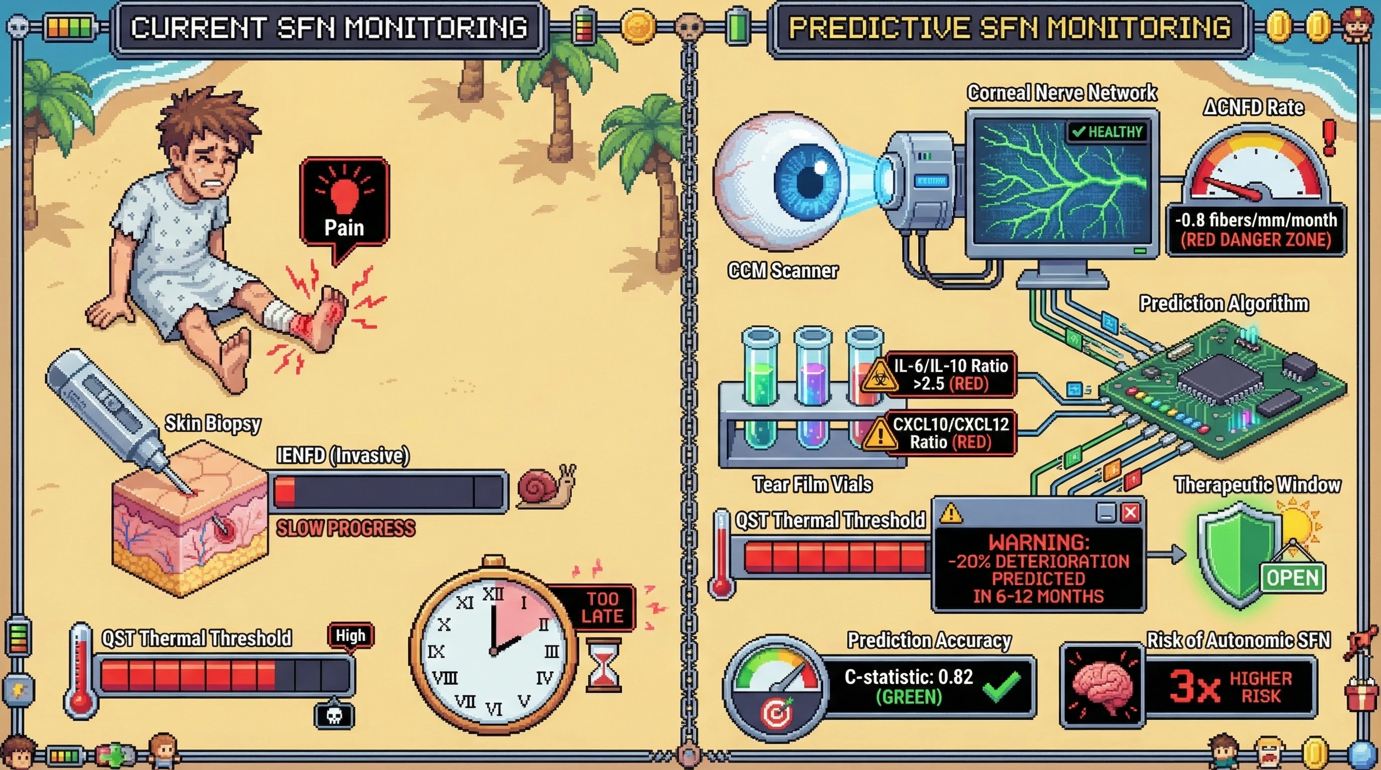
Small fiber neuropathy (SFN) affects 40–60% of primary Sjögren syndrome (pSS) patients, often preceding sicca symptoms and evading detection by conventional nerve conduction studies. Current diagnosis relies on skin punch biopsy (intraepidermal nerve fiber density, IENFD) — an invasive, non-repeatable procedure ill-suited for longitudinal monitoring. Corneal confocal microscopy (CCM) quantifies sub-basal corneal nerve fiber density (CNFD), branch density (CNBD), and fiber length (CNFL) non-invasively in minutes, and the cornea shares trigeminal innervation patterns homologous to peripheral small fibers.
Hypothesis
Serial CCM-derived CNFD decline rate (ΔCNFD/month), when combined with tear film inflammatory cytokine ratios (IL-6/IL-10, CXCL10/CXCL12), predicts systemic SFN progression — defined as ≥20% worsening in quantitative sensory testing (QST) thermal thresholds — 6–12 months before QST deterioration becomes clinically detectable in pSS patients.
Mechanistic Rationale
Corneal nerves undergo Wallerian-like degeneration driven by the same dorsal root ganglion (DRG) T-cell infiltration and anti-neuronal antibody-mediated damage that causes systemic SFN in pSS. The cornea, being the most densely innervated tissue in the body (~7,000 nociceptors/mm²), amplifies early neurodegenerative signals before they manifest in less densely innervated dermatomes. Tear film cytokine shifts from reparative (IL-10, CXCL12) to pro-inflammatory (IL-6, CXCL10) profiles reflect local neuroinflammatory escalation that parallels systemic DRG inflammation.
Testable Predictions
- Primary: pSS patients with ΔCNFD < −0.8 fibers/mm/month over 3 consecutive visits will develop ≥20% QST thermal threshold worsening within 12 months (HR >3.0, 95% CI excluding 1.0)
- Secondary: Adding tear IL-6/IL-10 ratio >2.5 to the CNFD slope model improves C-statistic from ~0.72 (CNFD alone) to >0.82
- Negative control: CNFD decline rate will NOT predict large fiber neuropathy (NCS-confirmed) progression, confirming specificity for small fiber pathology
- Dose-response: Patients in the fastest CNFD decline tertile will have 3× higher risk of developing autonomic SFN symptoms (orthostatic intolerance, gastroparesis) versus the slowest tertile
Proposed Study Design
Prospective cohort, n=150 pSS (ACR/EULAR 2016 criteria), quarterly CCM + tear cytokine panels + semiannual QST + annual skin biopsy (IENFD as gold standard). 24-month follow-up. Time-dependent Cox regression with CNFD slope as continuous time-varying covariate. Bonferroni correction for 4 co-primary/secondary endpoints (α=0.0125).
Limitations
- CCM requires specialized ophthalmic equipment (Heidelberg Retina Tomograph III), limiting multicenter feasibility
- Tear cytokine panels lack standardization across laboratories
- Concomitant dry eye disease may cause corneal nerve changes independent of systemic SFN
- QST is operator-dependent and subject to patient cooperation bias
- 150 patients may be underpowered for subgroup analyses (autonomic SFN)
- Cannot distinguish anti-neuronal antibody-mediated from ischemic SFN mechanisms
Clinical Significance
If validated, serial CCM would replace invasive skin biopsies for SFN monitoring in pSS, enabling non-invasive, repeatable tracking at routine ophthalmology visits. Early detection of SFN progression 6–12 months before clinical worsening opens a therapeutic window for neuroprotective interventions (IVIG, rituximab) before irreversible axonal loss. The combined CCM + tear cytokine model could be integrated into existing ESSDAI frameworks as a quantitative neurological domain score.
LES AI • DeSci Rheumatology